Radiation Doses from Computed Tomography of The Brain Based on Head Sizes in A Diagnostic Center in Northern Nigeria

Authors

  • Muhamed Kabeer Sulayman Department of Physics, Faculty of Science, Universiti Putra Malaysia, Serdang 43400, Selangor, Malaysia
  • Ahmad Shamsudeen Aliyu Department of Radiology, Usmanu Danfodiyo University Sokoto, 840004 Sokoto State, Nigeria
  • Izdihar Kamal Department of Physics, Faculty of Science, Universiti Putra Malaysia, Serdang 43400, Selangor, Malaysia
  • Bishir Umar Department of Environmental Health technology, College of Liberal Studies, Hassan Usman Katsina Polytechnic, 2052, Katsina, Nigeria
  • Faruk Kabeer Umar Department of Radiology, Usmanu Danfodiyo University Teaching Hospital Sokoto, 840004 Sokoto, Nigeria
  • Mohamad Asmawi Mohamad Arifin Department of Physics, Faculty of Science, Universiti Putra Malaysia, Serdang 43400, Selangor, Malaysia
  • Muhammad Khalis Abdulkarim Department of Physics, Faculty of Science, Universiti Putra Malaysia, Serdang 43400, Selangor, Malaysia

DOI:

https://doi.org/10.37934/sijphpc.4.1.5258

Keywords:

CT brain, effective dose, effective diameter, radiation dose

Abstract

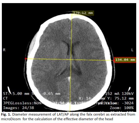
The study aims to evaluate and assess radiation dose and its relationship with the effective diameter of the head for CT brain examinations conducted using a 16 multi-slice CT scanning machine. Data from 30 patients who had CT brain examinations done at our facility was retrospectively collected in the month of May 2024. Data, including the volume Computed Tomography Dose Index (CTDIvol) value, dose-length product (DLP) value, scan range and the head diameter of the patient measured in the Antero-posterior (AP) and Lateral (LAT) orientations, were documented in a standardized format for analysis. The effective dosage E was then calculated. The mean ± S.D of E for brain CT was 2.5 ± 0.5 mSv. The mean ± S.D for the Effective Diameter DEFF was documented as 159.9 ± 8.7. The correlation (R2) between the E and DEFF showed 0.3315 as its values, indicating a positive correlation. The radiation increases according to the increase in head diameter. This study demonstrates that radiation exposures from CT brain scans may rely on the size of the head. Consequently, additional safety protocols should be implemented for the type of examination so as to reduce the potential risks linked to CT scans.

Author Biographies

Muhamed Kabeer Sulayman, Department of Physics, Faculty of Science, Universiti Putra Malaysia, Serdang 43400, Selangor, Malaysia

ibnsualeh@gmail.com

Ahmad Shamsudeen Aliyu, Department of Radiology, Usmanu Danfodiyo University Sokoto, 840004 Sokoto State, Nigeria

shamsudeen.aliyu@gmail.com

Izdihar Kamal, Department of Physics, Faculty of Science, Universiti Putra Malaysia, Serdang 43400, Selangor, Malaysia

izdiharkamal@gmail.com

Bishir Umar, Department of Environmental Health technology, College of Liberal Studies, Hassan Usman Katsina Polytechnic, 2052, Katsina, Nigeria

bishirumarelladan@gmail.com

Faruk Kabeer Umar, Department of Radiology, Usmanu Danfodiyo University Teaching Hospital Sokoto, 840004 Sokoto, Nigeria

faroukkbr@gmail.com

Mohamad Asmawi Mohamad Arifin, Department of Physics, Faculty of Science, Universiti Putra Malaysia, Serdang 43400, Selangor, Malaysia

mawie1107@gmail.com

Muhammad Khalis Abdulkarim, Department of Physics, Faculty of Science, Universiti Putra Malaysia, Serdang 43400, Selangor, Malaysia

mkhalis@gmail.com

Downloads

Published

2025-06-30

Issue

Section

Articles