
DiRetina: Anatomically Guided Hybrid Deep Learning for Glaucoma Detection from Retinal Fundus Images

Authors

  • Mohammad Loqman Hakeem Faculty of Information Sciences and Engineering, Management and Science University, Malaysia
  • Muhammad Irsyad Abdullah Faculty of Information Sciences and Engineering, Management and Science University, Malaysia
  • Ahgalya Subbiah Software Engineering and Digital Innovation Centre, Management and Science University, Malaysia

Keywords:

Glaucoma detection, retinal fundus imaging, deep learning, optic nerve head, hybrid feature fusion, medical artificial intelligence

Abstract

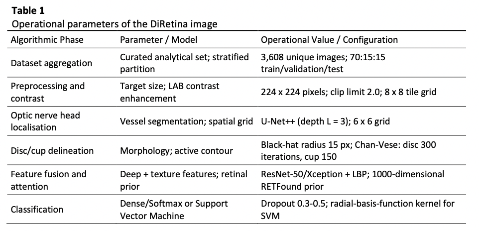
Glaucoma is a major cause of irreversible visual impairment, and early screening remains difficult because disease progression may be asymptomatic while manual fundus assessment depends on specialist availability and observer judgement. This study develops DiRetina, a multi-stage artificial intelligence framework for automated glaucoma screening from color retinal fundus photographs, with the objective of improving robustness under heterogeneous acquisition conditions while retaining interpretable anatomical markers. A curated analytical dataset of 3,608 unique images was formed from seven public retinal repositories and stratified into training, validation, and test subsets using a 70:15:15:15 allocation. The pipeline applies contrast normalization, vessel-guided optic nerve head localization using U-Net++, Chan-Vese active-contour delineation of the optic disc and cup, vertical cup-to-disc ratio extraction, and hybrid feature fusion combining Local Binary Patterns with pretrained convolutional representations. A knowledge-enhanced attention module incorporates frozen retinal foundation-model embeddings to reduce task-irrelevant background influence. Baseline ResNet-50, VGG-16, and Xception models achieved 86.70%-88.46% accuracy, with specificity between 78.62% and 82.63%. The optimized Xception configuration reached 95.07% accuracy and an area under the receiver operating characteristic curve of 0.9880, while the hybrid feature-Support Vector Machine configuration achieved 96.4% accuracy, 95.1% sensitivity, 97.2% specificity, and an area under the curve of 0.981. Reported one-way analysis of variance results indicated significant performance differences among model variants at p < 0.05. The findings support a structured screening strategy that combines anatomical localization, handcrafted texture descriptors, and deep representations to improve diagnostic consistency across heterogeneous fundus images. Prospective clinical validation remains necessary before deployment.

Author Biographies

Mohammad Loqman Hakeem, Faculty of Information Sciences and Engineering, Management and Science University, Malaysia

mohammadloqman02@gmail.com

Muhammad Irsyad Abdullah, Faculty of Information Sciences and Engineering, Management and Science University, Malaysia

irsyad@msu.edu.my

Ahgalya Subbiah, Software Engineering and Digital Innovation Centre, Management and Science University, Malaysia

ahgalya_subbiah@msu.edu.my

Downloads

Published

2026-10-11

Issue

Section

Articles